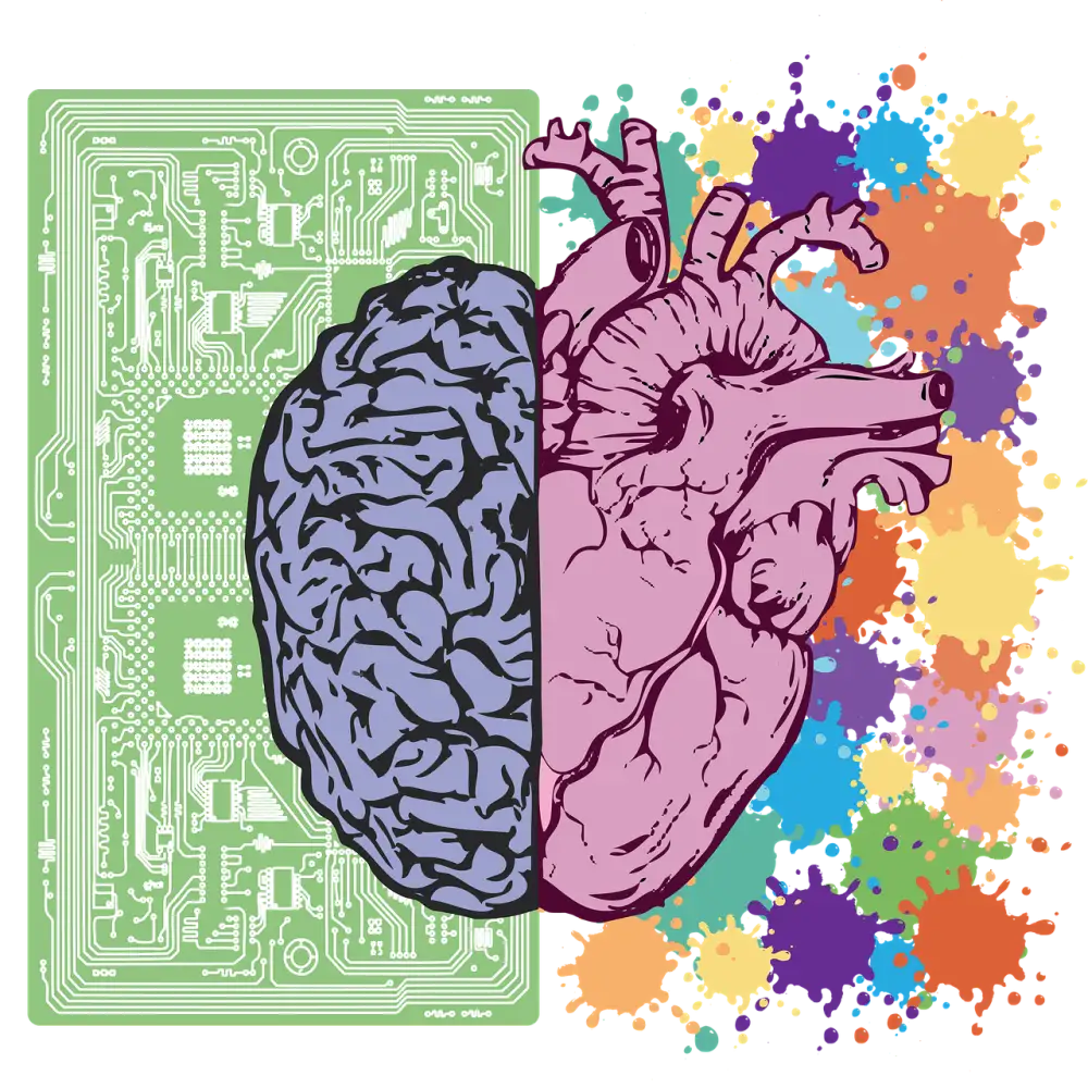
The Intriguing Anatomy: Unveiling the Human Brain Picture for Better Health Understanding

The human brain is a complex organ that plays a crucial role in our daily functioning. Visualizing the brain through imaging techniques is essential for medical diagnosis and research purposes. By examining detailed images of the brain, healthcare professionals can better understand its structure and identify any abnormalities or injuries. This visual representation allows for more accurate diagnoses and treatment plans, leading to improved patient outcomes.
Anatomy of the Human Brain:
The human brain, a complex organ weighing about 3 pounds, is divided into different regions visible in brain imaging. The cerebrum, the largest part responsible for thinking and voluntary movements, dominates the image. The cerebellum, located at the back of the brain, controls balance and coordination. The brainstem regulates basic life functions like breathing and heartbeat. The limbic system, involved in emotions and memory, is also depicted in the image. Each region plays a crucial role in our daily functioning and overall well-being.
Common Medical Uses of Brain Imaging:
Brain imaging plays a crucial role in diagnosing various conditions such as tumors, strokes, and traumatic brain injuries. By providing detailed images of the brain's structure, imaging techniques like MRI and CT scans help healthcare professionals identify abnormalities and plan appropriate treatment strategies. Moreover, brain imaging is instrumental in understanding complex neurological disorders like Alzheimer's and Parkinson's disease by revealing changes in brain structure and function associated with these conditions. Early detection through brain imaging can lead to timely interventions and improved patient outcomes.
Research Applications of Brain Imaging:
Brain imaging plays a crucial role in advancing neuroscience research by providing valuable insights into the inner workings of the human brain. Through techniques like functional magnetic resonance imaging (fMRI), researchers can map brain activity in real-time, allowing for a better understanding of cognitive functions such as memory, attention, and decision-making. These images help scientists uncover neural pathways involved in various processes, paving the way for innovative treatments and interventions for neurological conditions.
Future Trends in Brain Imaging:
Advancements in brain imaging technology are revolutionizing the field of neuroscience. Cutting-edge techniques such as diffusion tensor imaging (DTI) and magnetoencephalography (MEG) offer unprecedented insights into the brain's structure and function. These innovations enable researchers to map neural pathways with greater precision and study real-time brain activity. As technology continues to evolve, we can anticipate even more sophisticated imaging tools that provide detailed information for personalized medicine, leading to tailored treatment strategies for various neurological conditions.
Published: 11. 04. 2024
Category: Health


